The plantar fascia is a band of thick connective tissue on the bottom of the foot which supports the arch. It extends from calcaneus to the metatarsal bones like a cable connecting the front and back portions of the foot. Abnormalities of the plantar fascia include plantar fasciitis (an inflammatory process), fibroma (a benign fibrous soft tisse mass) and tear ( possibly related to previous cortisone injection). The plantar fascia serves a major role in the biomechanics of the foot. In the setting of a plantar fascia tear, the arch loses support leading to collapse.
A tear of the plantar fascia can be due to direct trauma, such as a sharp blow or jumping. As mentioned above, rupture of the plantar fascia can be related to previous cortisone injection. In addition, certain antibiotics like Cipro or levaquin are associated with plantar fascia tears.
Symptoms of a tear of the plantar fascia include localized heel pain, swlling, bruising, limping, and a sensation of the arch falling. Some patients hear apop when they tear the plantar fascia.
Treatment of a plantar fascia tear begins with immobilization, possibly in a cast or walking boot. Steroid injections and certain NSAIDs can help reduce symptoms, but they can also delay healing. Surgery may be necessary in chronic cases and can be quite successful.
MRI scanning is particularly useful in diagnosing tears of the plantar fascia and differentiating them from plantar fasciitis or a fibroma. Sometimes a tendon injury or stress fracture can mimic a tear of the plantar fascia. MRI can easily distinguish these entities from a plantar fascia tear.
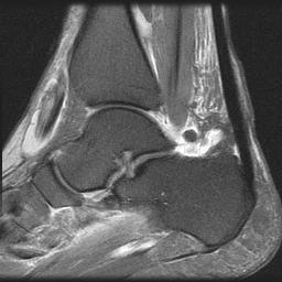 Normal Plantar Fascia
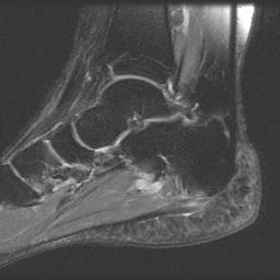 Plantar Fascia and tear
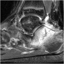 Stress Fracture
Plantar Fascia in the media - February 2013
Gasol to undergo MRI Wednesday in Boston