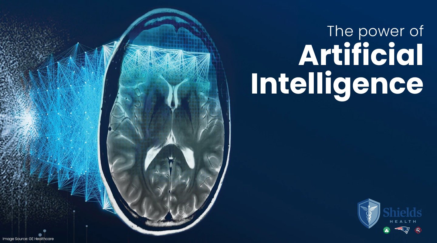
Posted by Siobhan Quinn
As the New England Patriots place Stephen Gostkowski on the injured reserve due to a torn quadricep & Shayne Graham steps in as the #1 kicker, folks are asking what does a tear look like on an MRI?
So here you have an example of a quadricep tear (left image) and a normal quadricep (no tear on the right):
Posted by Steve Sweriduk
The problem:
The American Cancer Society (ASC) estimates there will be 222,520 new cases of lung cancer this year alone. These new cases account for 15%+/- of cancer diagnoses annually. In the United States, lung cancer is the most common cause of cancer related deaths for both men and women with cigarette smoking being the number one cause for developing lung cancer.
Screening for Lung Cancer:
Based upon the findings of a study from the National Lung
Screening Trial (NLST), CT screening for lung cancer had been shown to be effective in reducing mortality from lung cancer. Since 2002, 53,000 men and women with at least a 30 “pack year” history of smoking were enrolled in the trial. Patients were randomly assigned to annual screening with either a low-dose (i.e., less radiation) CT scan or routine chest X-ray. The results demonstrate that annual screening with low-dose chest CT in heavy smokers, past or present, is far superior to conventional x-ray screening in detecting lung cancer and resulted in 20% fewer deaths from lung cancer. The findings were so impressive that the study was halted prematurely. An additional benefit of the study was a 7% reduction in all causes of death in patients screened with low dose chest CT. More detailed information can be found on the National Cancer Institute website: www.cancer.gov .
While it is clear from the NLST study that screening former and current heavy smokers increases survival from lung cancer, it remains unclear if light or moderate smokers will achieve similar benefits from CT screening.
The cost of a low-dose chest CT is approximately $350-450 at an out-patient imaging facility and the radiation dose is similar to that of a mammogram.
Conclusion:
This is a promising development. It is now clear low dose chest CT screening will play a vital role in the fight against lung cancer. Yet the most important step any of us can take in the battle against lung cancer is to quit smoking or never start smoking. Although quitting smoking reduces but does not eliminate the risk of developing lung cancer it is a critical step. For those who have previously had lung cancer, quitting smoking reduces the risk of developing a second lung cancer.
Clearly, the benefits of low-dose chest CT screening for diagnosing a treatable lung cancer outweigh the risks associated with radiation exposure for heavy smokers. But the best thing you can do in the fight against lung cancer is to quit smoking or never start. For those who need help to quit smoking contact your primary care physician or check some online resources such as: www.smokefree.gov; and the American Cancer Society www.cancer.org.
Posted by Carmel Shields
Check out Gardiner Harris' article in the New York Times: "CT Scans Cut Lung Cancer Deaths, Study Finds." Mr. Harris reports on a recent study that finds annual CT scans for 'heavy' smokers can aid in reducing deaths by lung cancer by 20%!
Unfortunately, Medicare and health insurers do not (currently) cover the cost of CT lung screening. This means the individual will be out-of-pocket approximately $350 to $450 for a scan at an out-patient imaging center versus a scan at an academic medical center or large hospital system where charges, for the same scan-quality-read & equiment, run as high as $1865 for the scan PLUS $395 for the professional read by the radiologist.
To read more from Gardiner Harris and the New York Times go to: http://www.nytimes.com/2010/11/05/health/research/05cancer.html
Posted by Steve Sweriduk
Virtual colonoscopy (VC) is an examination of the large intestine and rectum involving a CT scan after the introduction of gas into the colon. Another name for the procedure is CT colonography.
The primary objective of the exam is to screen for abnormalities of the large bowel including polyps or cancer.
Why have a VC?
Colorectal cancer is the second most common cause of cancer related deaths in the U.S. after lung cancer. Finding and removing colon polyps before they can become cancerous can reduce the incidence of colon cancer dramatically. Current recommendations for colon screening include a routine colonoscopy every 10 years beginning at age 50, with earlier and or more frequent screening in other situations such as family history of colorectal cancer and certain types of polyps. Virtual colonoscopy every 5 years is a good alternative for colon screening.
Process
Both conventional and virtual colonoscopies require you to take a laxative or other agent to make sure that the colon is free of stool during the examination. This is necessary to increase the accuracy for detecting abnormalities. Unlike conventional colonoscopy, Virtual Colonoscopy requires no intravenous sedation during the procedure. This allows the patient to swiftly return to usual activities immediately after the procedure with little “post op” recovery.
Cost
The cost of virtual colonoscopy is half that of a conventional colonoscopy. There are “virtually” no complications associated with a virtual colonoscopy. Conventional colonoscopy is also a very safe procedure, but does have some potential complications, including risks associated with sedation and the remote chance of bowel perforation. It can be difficult to evaluate the entire bowel with conventional colonoscopy, not so with VC.
Advantages and Disadvantages
VC is better tolerated than conventional colonoscopy, with less discomfort. The entire abdomen and pelvis are imaged with CT, not just the bowel lining. This may help detect unsuspected lesions. Disadvantages of VC include radiation exposure and some limitation in identifying polyps less than 1 cm in size. However, polyps less than 1cm are usually insignificant. Another disadvantage of VC is that it is a purely diagnostic study. If a significant polyp is found, it will need to be removed by conventional colonoscopy or through surgery. The flip side of the argument is that VC can certainly eliminate unnecessary colonoscopies, the discomfort of traditional colonoscopy procedures and reduce overall health care expenses.
Additional Resources
For more information you may want to go to the following websites: www.nih.gov (National Institutes of Health); www.nci.gov (National Cancer Institute); www.rsna.org (Radiology Association of North America), to name a few.
Posted by Steve Sweriduk
What does a broken collarbone /clavicle look like on a CT scan?
According to news reports, Tony Romo of the Dallas Cowboys had a CT scan showing a broken left clavicle. At this point it appears he will not need surgery but will be off the field for 6 to 8 weeks.
Clavicle fundamentals
The clavicle (collarbone) connects the shoulder to the chest wall. Fractures can result from either an indirect trauma like a fall to an outstretched arm or due to a direct hit to the collarbone.
Sports with a high probability of direct or indirect trauma from falls or contact include football, hockey, and skiing. The clavicle, or collarbone, rests over very important structures: the subclavian artery and vein are the major circulatory structures supplying the arm with blood. The brachial plexus are the nerves coming out of the neck and going to the arm. Clavicle fractures can, but rarely do, cause injury to these structures.
Symptoms
As with any like fracture acute clavicle or shoulder pain, swelling and bruising are the most common symptoms along with pain when moving the arm.
What to do
Immediately after injury or impact the arm should be immobilized in a sling for comfort. To reduce swelling and pain, ice may be applied and anti-inflammatory medication used if appropriate.
Treatment options
Over 90 percent of such fractures heal without surgery. Two common options are use of a sling or a splint (i.e., figure 8 splint). Both are meant to keep the fragments still throughout the healing process of approximately 6 to 8 weeks. As your bones mend, you can start limited motion exercises to help prevent a frozen shoulder.
Most fractures do not require surgery; however, surgery may be appropriate if the collarbone is widely separated. Surgery is also recommended when nerve vessel injury occurs or if the fracture has not healed properly. If the fracture brakes through the skin surgery may be appropriate to minimize the risk of infection.
Posted by Steve Sweriduk
Soccer, football and marathon training are in full swing and we are seeing an infux of patients with an injury to the hamstring.
What is the Hamstring?
The hamstring is a group of muscles located on the back of the thigh. The hamstring muscles allow the leg to bend and extend.
Injuries to the Hamstring
Hamstring injuries can occur in a number of ways. Common examples include:
- muscle fatigue
- direct hit to the thigh
- insufficient warm-up or conditioning
- insufficient rest and recovery of a previous hamstring injury
To aid in the recovery of a hamstring injury remember RICE - rest, ice, compression and elevation.
Here are two MRI images of the hamstring - a normal (non injured) hamstring and an injured hamstring.
To stay in the game remember to take time to allow the injury to heal.
Posted by Steve Sweriduk
Marc Savard and Don Hasselback have raised awareness of concussions and the toll head trauma can take on the individual, as an athlete and in everyday life.
A concussion results from head trauma and can result in
confusion, memory loss, and loss of consciousness. Concussions are a common injury, with over 1 million cases reported per year in the United States alone. Sports commonly associated with concussion include football, soccer, hockey, boxing, lacrosse and of course, hockey. As many as 10% of college and 20% of high school football players will suffer from a concussion annually.
What are the Symptoms?
Symptoms of a concussion include headache, dizziness, nausea and or vomiting, slurred or incoherent speech, and loss of balance and coordination. Symptoms may develop immediately after injury or develop over a period minutes to hours.
Grading a Concussion
There are 3 grades of concussion. Grade 1 is the least severe and involves “transient” confusion lasting less than 15 minutes and without loss of consciousness. Grade 2 is more significant with symptoms of confusion or mental status changes lasting more than 15 minutes but without loss of consciousness. Any concussion resulting in loss of consciousness is categorized as Grade 3. The more information you have about your athlete prior to injury the better. For example, pre-season (pre-injury) baseline testing helps the physician gauge and grade the extent of injury.
Returning to the Sport
There are several well-published guidelines to help physicians determine when it is safe for athletes to return to play after a concussion. The guidelines are somewhat controversial, but all agree no player should return to sports until symptoms are completely resolved. Patients with symptoms lasting more than 15 minutes or who exhibit memory loss should be restricted from sports participation for at least 1 week. Any player who loses consciousness should be evaluated in the emergency room by medical personnel. For a Grade 3 concussion, some guidelines suggest that the athlete should not return to play for a minimum of 1 month or longer. If a player sustains multiple concussions, some suggest that the season should be terminated. It is important that players, parents, coaches, trainers and physicians understand the serious nature of concussions.
Diagnostic Testing for Concussion
Imaging of the brain with CT or MRI is usually not indicated unless the patient’s symptoms are severe or if there has been loss of consciousness. MRI and CT scans of patient’s with concussion are usually normal in the acute phase, although on rare occasions edema and bleeding in or around the brain can be seen. Follow-up CT and MRI scans can show brain atrophy. Brain function measurements using MRI can be abnormal, corresponding to decreased performance on neuropsychological testing.
Autopsy studies of former football players and boxers have shown brain injury due to multiple and repetitive head trauma resulting in chronic traumatic encephalopathy with accumulation of large amounts of tau protein in areas of the brain responsible for mood, emotion and executive functioning. These same proteins are found in patients with Alzheimer’s disease.
Safety Gear and Education
Protective gear such as helmets and mouth guards can reduce the incidence of concussion, but cannot completely prevent against it. Education on the risks associated with concussion is essential.
Going Forward
Prompt diagnosis, appropriate restrictions, and treatment may reduce the risk of further injury, but the long term consequences are not fully known.
Concussion is a very serious and potentially life changing/threatening disorder. To assist in the medical evaluation secure a baseline of your athlete and be sure he/she wears appropriate safety gear to minimize impact and trauma.
Posted by Carmel Shields
A recently released study from Sweden is fostering debate over what age women should get mammograms.
This most recent study suggests that mammograms, a breast cancer screening test, can lower the risk of dying of the disease by 26 percent or more for women in their 40s. These findings conflict with the U.S. panel that recommended against routine screening before the age of 50.
Its timely to post an abbreviated article by Alan Semine, MD, Chief of Breast Imaging, Shields MRI, and Chief of Breast Imaging for Newton-Wellesley Hospital; and Clincila Professor of Radiology, Tufts University School of Medicine, on the issue.
Why the U.S. Preventive Services Task Force is Wrong
The screening mammography recommendations of the United States Preventive Services Task Force, (USPSTF) published recently in the Annals of Internal Medicine and widely reported by the press, were poorly stated and misleading. In fact, the USPSTF based its dubious conclusions on existing data that it reinterpreted. The recommendations have been challenged by organizations including the American Cancer Society, the American College of Radiology, the American Society of Breast Surgeons, the Society of Breast Imaging, the American Society of Clinical Oncology and many others.
The USPSTF recommended against routine screening for women 40 to 49 years of age. Yet the task force acknowledged that mammography in this age group does save lives. This fact is not disputable based on extensive long term studies. Its recommendation was based on its own conservative risk benefit valuation.
The USPSTF rendered a verdict that screening 1339 women in their 50s to save one life makes screening worthwhile in that age group. But because it is necessary to screen 1904 women in their 40s to save one life, then screening is not worthwhile. Such valuation in the face of relatively comparable numbers is not justified. In fact, there is excellent data demonstrating even better survival with mammography screening. The task force recommended screening for those of high risk yet ignored the fact that 70% of breast cancers develop in women without family history. The financial cost of treatment for advanced breast cancer is also far greater than for early stage cancer.
The next recommendation of the task force was to screen women over fifty every other year rather than annually. They acknowledged the obvious: that the cancers detected, on average, would be more advanced but they emphasized that the total number of cancers detected would be the same with fewer questionable findings. This position represents a serious lack of understanding that the most important objective of screening mammography is to detect breast cancer at the earliest possible stage to get the best chance of cure. Meanwhile, the task force also suggested that screening can be stopped after age 74. Yet, it is well established that the incidence of breast cancer continues to increase with age.
The recommendations of the USPSTF are not based on new data but rather on a reinterpretation of selected studies. They reached a conclusion that differs markedly from the position of national organizations as well as prior recommendations of the USPSTF. The members of the task force do not represent a new standard in medical wisdom. The publicity they have received threatens to undermine decades of effort educating women about the importance of early detection to combat breast cancer.
Mammography does have limitations and it is essential that we pursue every opportunity to improve its effectiveness as well as explore alternative methods of detection and treatment.
We must continue to urge women to begin screening mammography at age 40 and encourage them to talk with their doctors to ask any questions they have about mammography and self-examination. Alan Semine, MD
Posted by Siobhan Quinn
To plan for your MRI, CT or PET scan, it may help to better undertand the how short or long it takes for an MRI, CT or PET scan.
The scan time, the time you are actually in the machine as it acquires images, depends on a number of factors. Namely, the type of test, area of the body being imaged and if a contrast agent is recommended. Prior to the actual scan, patient registration and meeting with the technologist takes approximately 30 minutes.
Here are typical timelines for your scan:
MRI (magnetic resonance imaging) scan times will take 30 to 40 minutes for the majority of scans. That is, brain, knee, spine and shoulder. However, if the study requires a contrast agent your scan time can be 50-60 minutes. Prostate and breast MRI scans take 60 to 80 minutes.
CT (computerized tomography) scan times are often shorter than those for an MRI. A CT scan without contrast (head, sinus, orbits, abdomen, pelvis, cervical, thoracic, lumbar and dental scans) take approximately 15 minutes. CT studies with contrast of the chest, pelvis, head or neck take approximately 30 minutes. Whereas, aortic contrast studies may take up to 45 minutes. Most cardiac CT studies will take 60 minutes.
PET (positron emission tomography) scan times take longer than either MRI or CT scans because the injection of a radioisotope take 45 to 1.5 hours to travel through the body. The typical scan (or image acquisition) time for brain and oncology related scans is 30 to 60 minutes. Consequently, a complete PET scan customarily takes 2 hours. Sometimes, an additional 20 minutes of imaging time is necessary for scans related to melanoma and sarcoma.
I hope these guidelines help you plan ahead for your scan.
Would you like to share your experience?
Posted by Christine Mavilia
Before launching a Wellness Program decide what you are trying to achieve and set realistic timelines for achieving a return on your investment.
According to the Wellness Council of America, for every $1 investment in Wellness Programs you can expect to save $3 in health care costs.
But how quickly?
Well, the return is dependent on the type of program adopted. We recommend you consider the following:
- Recruit and target employees who are driving your health
care costs upward. Bring screenings in-house and incentivize employees to participate. Simple steps like blood pressure checks, cholesterol and glucose screening give employees information to begin to make healthier choices.
- Partner with the disease management departments of your health insurance carrier. This means your carrier will reach out to specific employees based on claims data and provide them with counseling or group support based on their specific disease or situation. This type of coaching can often bring about a faster return on your “wellness” spend.
- Health Plan Design – Employees are more accountable for their own health when they are keenly aware of the cost. Instituting a high deductible plan helps employees recognize the price of lifestyle choices. In a consumer driven plan, employees are price sensitive and will research cost. For example, pricing generic and mail order medications or contact lenses because of the dollar difference. At bottom, realizing a return on investment using plan design can take several plan cycles.
- Exercise, Nutrition and Point-Based Programs - Award points for different lifestyle choices that translate into (health related) gifts are common. They should be low cost, easy to implement and create excitement and competition within your organization. The downside is those that are quick to join are often the most healthy! But if you have one champion success is more likely.
- Utilize free resources. Reach out to your EAP, health plan and community resources (such as smoking cessation programs) to lower your overall financial investment, while still providing a robust plan.
Wellness programs are a hot topic and success is dependent on actively managing the overall health of your employee population and rising premiums.
This means get started now - delay is not an option!